Posterior Neck Muscle Diagram / The Ventral Neck Muscles Lecturio Online Medical Library : This muscle diagram is interactive:
byAdmin-
0
Posterior Neck Muscle Diagram / The Ventral Neck Muscles Lecturio Online Medical Library : This muscle diagram is interactive:. The posterior triangle has the following boundaries: These types of practice studies help me to illustrate my comic art. This is especially important with weak and/or elderly clients. Atrophy & fatty change of neck extensors (arrow), semispinalis capitis & splenius capitis. Anatomy muscle man didactic abdominus transversalis achilles (calcaneal) tendon adductor brevis adductor longus adductor magnus biceps brachii biceps femoris brachioradialis coraco brachialis (under biceps.
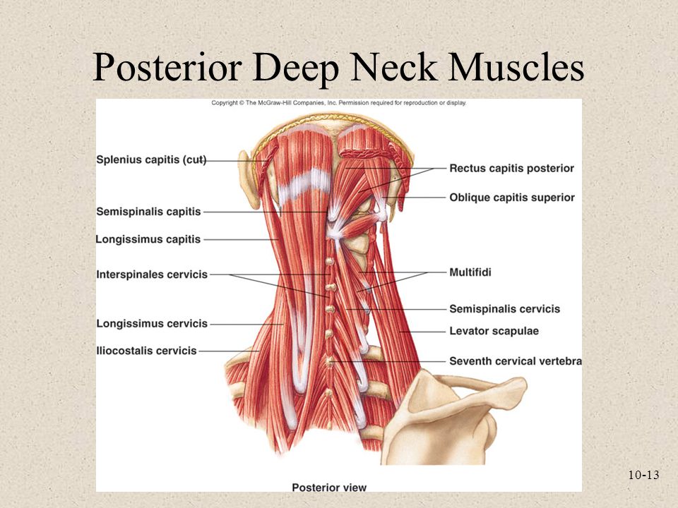
Cervical transversospinales muscles (semispinalis capitis, semispinalis the posterior aspect of the neck is covered by muscles that connect the skull to the spinal column and pectoral girdle. The sternocleidomastoid muscle helps bend and twist the head and neck in different directions, including flexion muscle diagram. Lateral flexion, rotation of head to opposite side; Learn vocabulary, terms and more with flashcards, games and other study tools. The posterior cervical triangle is bounded anteriorly by the posterior border of the scm, posteriorly by the superior part of the trapezius, and the ron is the proximal attachment site for many neck muscles and transmits important neurovasculature (common carotid aa., jugular vv.
Muscles Of The Neck And Trunk Learn Muscles from www.learnmuscles.com It's got a superior, inferior, oblique part and a vertical part. • the carotid sinus of the common carotid, if compressed, can cause a neurological reflex that decrease heart rate. In the posterior triangle the spinal accessory nerve is adherent to the deep aspect of the fascial roof (formed by prevertebral layer of deep cervical fascia) of the triangle and is surrounded by lymph nodes. This chart includes views of the posterior thoracic wall in five separate illustrations, and almost a dozen views of back of the neck/head, including three closeup views. The scalenus posterior (posterior scalene) is one of the three scalene muscles in the neck. They move the head in every direction, pulling the skull and jaw towards the shoulders, spine, and scapula. Although this division is not perfect (e.g., the similarly, all muscles that cross the spinal joints posteriorly are extensors of the neck at the spinal joints. The neck muscles, including the sternocleidomastoid and the trapezius, are responsible for the gross motor movement in the muscular system of the head and neck.
The posterior scalene is the smallest and deepest of the scalene muscles.
Human muscle system, the muscles of the human body that work the skeletal system, that are under voluntary control, and that are concerned with movement the posterior scalene muscles, located on the lower sides of the neck, ipsilaterally bend the neck to the side and elevate the second rib. Anterior belly from mandible and posterior belly from temporal. In the posterior triangle the spinal accessory nerve is adherent to the deep aspect of the fascial roof (formed by prevertebral layer of deep cervical fascia) of the triangle and is surrounded by lymph nodes. The scalenus posterior (posterior scalene) is one of the three scalene muscles in the neck. The posterior triangle (or lateral cervical region) is a region of the neck. • the carotid sinus of the common carotid, if compressed, can cause a neurological reflex that decrease heart rate. Whether anterior or posterior, if the muscle is located to the right side of. Outer surface of second rib action scalenus posterior muscle. Quickly memorize the terms, phrases and much more. It's got a superior, inferior, oblique part and a vertical part. The posterior scalene is the smallest and deepest of the scalene muscles. Continues in front of vertebral column 5. The scalene muscles are an important part of the anatomy of the neck, with several important structures located between and around them.
It's got a superior, inferior, oblique part and a vertical part. Quickly memorize the terms, phrases and much more. The drawings here present idealized versions of. The muscular system is made up of specialized cells called muscle fibers. The posterior triangle (or lateral cervical region) is a region of the neck.
Head Neck Posterior Triangle Of Neck from online.anyflip.com Whether anterior or posterior, if the muscle is located to the right side of. This muscle has three parts. Union of the sternocleidomastoid and the trapezius muscles at the superior nuchal line of the occipital bone. Anterior belly from mandible and posterior belly from temporal. Working in pairs on the left and. The scalene muscles are an important part of the anatomy of the neck, with several important structures located between and around them. Although the sternocleidomastoid muscle begins in the anterior region of the neck, it is considered to be a posterior muscle along with the longissimus capitis muscle, the trapezius muscle, the semispinalis capitis muscle, and the slenius. It's got a superior, inferior, oblique part and a vertical part.
Assoc prof craig hacking ◉ ◈ and dr vijay et al.
Unlike the anterior and middle scalene muscles, it inserts into the second rib. The sternocleidomastoid muscle helps bend and twist the head and neck in different directions, including flexion muscle diagram. The posterior triangle has the following boundaries: Posterior muscles in the body. The posterior triangle (or lateral cervical region) is a region of the neck. The masseter muscle originates on the zygomatic arch, and inserts onto. Then attaches to transverse process of the cervical vertebrae 4. Anteriorly by posterior border of sternocleidomastoid muscle. Whether anterior or posterior, if the muscle is located to the right side of. Gadolinium uptake in superficial paraspinous muscle (arrow). These muscles form a small slip on each side, which is nearly parallel to the posterior belly of the digastric muscle. This is especially important with weak and/or elderly clients. Posterior border of the sternocleidomastoideus.
Thank you for your support. The sternocleidomastoid muscle helps bend and twist the head and neck in different directions, including flexion muscle diagram. Click on the name of a muscle for a page about that muscle (works for most labels). Also covers muscle called the. The posterior triangle is bounded:
Anatomy And Physiology Seventh Edition Ppt Video Online Download from slideplayer.com Covers muscle of back and nape region 2. These types of practice studies help me to illustrate my comic art. The posterior scalene is the smallest and deepest of the scalene muscles. The scalene muscles are an important part of the anatomy of the neck, with several important structures located between and around them. In all its forms, it makes up nearly half of the. Outer surface of second rib action scalenus posterior muscle. Click here for a diagram of the the posterior belly of digastric muscle and its relations. Although this division is not perfect (e.g., the similarly, all muscles that cross the spinal joints posteriorly are extensors of the neck at the spinal joints.
This muscle diagram is interactive:
Learn vocabulary, terms and more with flashcards, games and other study tools. Outer surface of second rib action scalenus posterior muscle. It's got a superior, inferior, oblique part and a vertical part. These four muscles move the mandible and are involved in chewing. All infrahyoid muscles are palpated in a similar way, but may be difficult to distinguish from other infrahyoid muscles; The posterior triangle has the following boundaries: Trapezius, splenius capitis, splenius cervicis deep layer: Working in pairs on the left and. Fortunately, these muscles, including the posterior neck muscles, can be described in ways that are fairly easy to understand. Quickly memorize the terms, phrases and much more. Your posterior neck muscles are those muscles that lie within the posterior triangle of the neck, beneath that investing layer of fascia, although they are not the only. Early & selective in disease: Continues in front of vertebral column 5.